Understanding a cow’s exterior is vital for effective livestock management, disease recognition, and overall animal well-being, as detailed in comprehensive anatomical guides.
A. Importance of Understanding Cow Anatomy
A solid grasp of bovine anatomy, particularly external features, is paramount for those involved in cattle care. For farmers, accurate identification of body parts aids in assessing overall health and detecting subtle changes indicative of illness or injury. Veterinarians rely heavily on anatomical knowledge for accurate diagnoses and effective treatment plans.
Furthermore, understanding external anatomy facilitates proper animal husbandry practices, including efficient handling, successful breeding programs, and appropriate nutritional management. Recognizing key landmarks assists in administering medications, performing procedures, and monitoring growth. Detailed guides, often available as PDFs, empower individuals to enhance herd management and promote animal welfare through informed observation and proactive care.
B. Scope of this Guide: Focusing on External Features
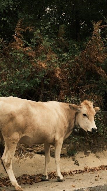
This guide concentrates specifically on the external anatomy of cattle – both cows and bulls – providing a detailed overview of visible body parts. We will systematically explore the head, encompassing the mouth, horns, eyes, ears, and nose, then proceed down the neck, body, forequarters, and hindquarters.
The reproductive organs, as viewed externally, and the tail will also be covered. Internal anatomy, while crucial, falls outside the scope of this document. Resources like downloadable PDFs often supplement visual learning, offering labeled diagrams and detailed descriptions of each external feature. This focused approach aims to equip readers with practical knowledge for identification and assessment of cattle.

II. The Head: A Detailed Examination
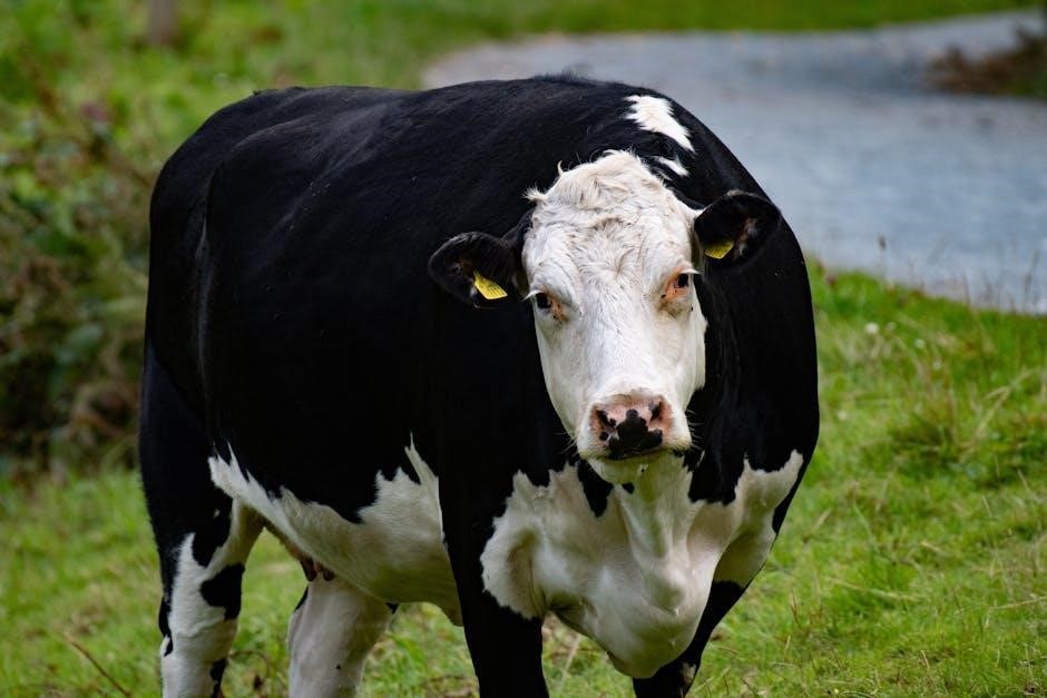
The bovine head showcases key features like the mouth, horns (if present), forehead, eyes, ears, and nose, all vital for function and identification.
A. Mouth and Lips
The cow’s mouth is specifically adapted for grazing, possessing a broad, tough dental pad instead of upper incisors. This pad works in conjunction with the lower incisors to grasp and tear grasses. The lips are thick and muscular, exhibiting considerable prehensility – meaning they can manipulate forage effectively. These lips play a crucial role in selecting desirable plants and efficiently gathering feed. Observing the mouth and lips can indicate dental health and potential feeding issues. Variations in lip length and shape can also be noted between different breeds. A healthy mouth is fundamental for proper nutrition and overall well-being in cattle, and careful examination is a key component of routine health assessments.
B. Horns (Presence, Shape, and Function)
Cattle horns are bony core structures covered in a keratin sheath, varying significantly in presence, shape, and size depending on breed and sex. While historically used for defense and establishing dominance within the herd, their function has diminished with modern farming practices. Horn shapes range from curved and pointed to shorter, blunter forms. Polled cattle, naturally hornless due to genetics, are increasingly common. Dehorning, the removal of horns, is practiced to prevent injury to animals and handlers. Horns can provide clues about an animal’s age and genetic lineage. Careful observation of horn development is important for assessing overall health and breed characteristics.
C. Forehead and Poll
The forehead, the area between the eyes and the base of the horns (if present), exhibits breed-specific variations in slope and width. The poll, situated directly behind the forehead, is a crucial anatomical landmark – a bony prominence where the head joins the neck. It’s a key assessment point for evaluating conformation and muscling. A well-defined poll indicates proper skeletal structure and influences the animal’s ability to flex its neck. Palpation of the poll can reveal subtle indicators of health or discomfort. Breed standards often specify ideal forehead and poll characteristics, impacting overall aesthetic appeal and functional efficiency.
D. Eyes and Eyelids
Cows possess large, horizontally-oriented eyes providing a wide field of vision, crucial for predator detection. Eyelids protect the eyes, featuring upper, lower, and nictitating (third) eyelids. The nictitating membrane sweeps across the eye for lubrication and debris removal. Observing eye clarity is vital; dullness or discharge signals potential illness. Breed variations exist in eye pigmentation – ranging from dark brown to lighter shades. Proper eyelid function ensures adequate tear distribution, maintaining corneal health. Regular examination of the eyes and surrounding tissues aids in early detection of infections or injuries, impacting overall animal welfare.
E. Ears and Their Role in Sensory Perception
Cows utilize their relatively large, mobile ears for acute auditory perception, essential for detecting sounds of predators or herd members. Ear positioning indicates mood; forward ears suggest alertness, while drooping ears may signal relaxation or illness. The pinna, the visible outer ear, funnels sound waves towards the inner ear. Cattle possess a wide range of hearing, though not as extensive as humans. Observing ear posture and cleanliness is crucial; flies and infections can cause irritation. Healthy ears contribute significantly to a cow’s awareness of its surroundings and social interactions within the herd.
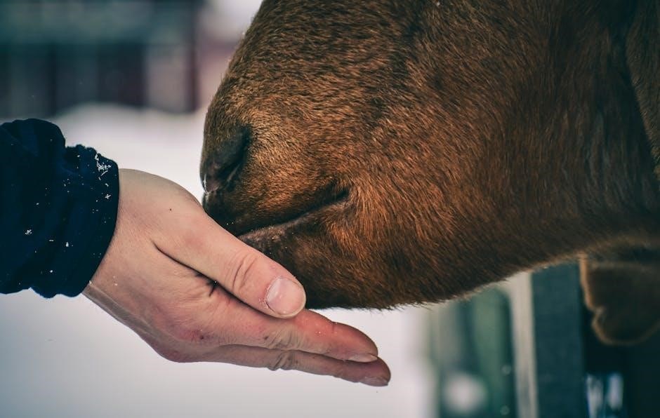
F. Nose and Nasal Cavity
The bovine nose, featuring a broad muzzle and nostrils, is critical for olfaction and respiration. Cows possess a highly developed sense of smell, used for identifying food, detecting predators, and recognizing other cattle. The nasal cavity warms and humidifies inhaled air, protecting the lungs. A moist nose generally indicates good health, while a dry or crusty nose may signal dehydration or illness. Observing nasal discharge is important; clear discharge is normal, but colored discharge suggests infection. The nose also plays a role in thermoregulation, aiding in cooling through respiration.

III. The Neck and its Key Features
The neck supports the head and contains vital structures like the dewlap and brisket, facilitating movement and housing essential muscles for grazing.
A. Dewlap (Function and Variation)
The dewlap, a prominent fold of skin hanging beneath the neck, exhibits considerable variation in size and development among cattle breeds. While its precise function isn’t fully understood, it’s believed to play a role in thermoregulation, potentially aiding in heat dissipation through increased surface area. It may also facilitate lymphatic drainage and serve as a protective cushion during social interactions, like neck wrestling in bulls.
Larger dewlaps are often observed in breeds adapted to warmer climates, suggesting a stronger thermoregulatory role. In bulls, a substantial dewlap can enhance their imposing appearance, potentially influencing dominance displays. The texture and looseness of the dewlap also vary, influenced by factors like age, nutritional status, and breed characteristics. Observing the dewlap can offer clues about a cow’s overall health and breed predisposition.
B. Brisket and Sternum
The brisket, located on the ventral midline of the chest, is a crucial area encompassing the sternum and associated cartilage. It forms the lower portion of the thorax, protecting vital organs like the heart and lungs. The sternum, comprised of several sternebrae, provides a rigid yet flexible structure. Palpating the brisket can reveal information about a cow’s respiratory health; swelling or sensitivity may indicate pneumonia or other chest infections.
Breed variations influence brisket conformation – some breeds exhibit a deeper, wider brisket than others. A well-developed brisket supports efficient respiration, particularly important for cattle engaged in strenuous activity. The angle of the ribs relative to the sternum also impacts breathing capacity. Assessing the brisket’s appearance and feel is a valuable component of a comprehensive physical examination.

IV. The Body/Barrel: Core Structure
The bovine body, or barrel, houses vital organs and is defined by ribs, flank, abdomen, back, and loin—essential for assessing overall health and conformation.
A. Ribs and Intercostal Spaces
The ribs form the foundational structure of the bovine body’s barrel, providing protection for vital organs like the heart and lungs. These curved bones articulate with the vertebral column posteriorly and the sternum anteriorly, creating the thoracic cavity. A mature bovine typically possesses 13 pairs of ribs, though variations can occur.
Externally, the intercostal spaces – the gaps between the ribs – are visible and palpable. These spaces allow for the expansion and contraction of the chest during respiration. Assessing the depth and symmetry of these spaces can offer insights into a cow’s respiratory health and overall body condition. Palpation can reveal abnormalities like bruising or swelling, potentially indicating underlying injuries or illness. Observing the ribcage’s conformation is crucial for evaluating a cow’s structural soundness and potential for efficient weight gain.
B. Flank and Abdomen
The flank region, located just behind the ribs, represents the lateral wall of the abdomen in cattle. It’s a crucial area for observing body condition and identifying potential health issues. A well-filled flank indicates good nutritional status, while a sunken flank can suggest weight loss or dehydration. The abdomen itself extends from the ribs to the pelvis and houses the digestive system and other vital organs.
Externally, the abdomen’s contour provides clues about the animal’s internal state. Distension can signal bloat or pregnancy, while asymmetry might indicate organ displacement or internal masses. The udder, prominent in female cattle, is located within the abdominal region. Careful observation of the flank and abdomen is essential for early detection of abnormalities and effective herd management, as detailed in bovine anatomy resources.
C. Back and Loin
The back and loin constitute the dorsal midline of the cow’s body, extending from the withers to the rump. The back’s topline reveals much about the animal’s conformation and muscling. A straight, level back is generally desirable, indicating structural soundness. The loin, situated just forward of the hips, is a key area for assessing muscle development and potential carcass quality.
Palpation of the loin can help determine the degree of muscling. A well-defined loin suggests a higher yield of valuable cuts of meat. The spinous processes of the vertebrae are visible externally and can be used to assess the animal’s overall health and posture. Observing the back and loin is crucial for breeders and producers aiming to improve herd genetics and meat production, as detailed in anatomical guides.
V. The Forequarters: Legs and Feet
The forequarters, including legs and feet, provide support and locomotion; their structure—shoulder, elbow, knee, and hoof—is essential for mobility and health.
A. Shoulder and Upper Arm
The shoulder represents the junction between the foreleg and the body, exhibiting significant muscular development crucial for locomotion and weight-bearing. Observing the shoulder’s conformation provides insights into the animal’s structural correctness and potential for efficient movement. The upper arm, extending from the shoulder to the elbow, houses substantial muscle mass responsible for propelling the forelimb forward.
Palpation of the shoulder and upper arm reveals key bony landmarks and muscle groups. Proper assessment of these areas aids in identifying potential injuries or conformational defects. A well-defined shoulder blade and a smoothly muscled upper arm indicate good conformation and athletic ability. Variations in muscle development can signal underlying health issues or breed-specific characteristics. Understanding this region is paramount for evaluating a cow’s overall physical condition and performance capabilities.
B. Elbow and Forearm
The elbow joint, a prominent bony projection, facilitates flexion and extension of the forelimb, enabling efficient locomotion. Careful examination of the elbow reveals its structural components and potential for conformational abnormalities. Extending distally from the elbow, the forearm comprises two bones – the radius and ulna – encased in robust musculature. This region provides leverage and support during movement.
Assessing the alignment and range of motion in the elbow is crucial for identifying lameness or joint issues. The forearm’s musculature contributes significantly to the cow’s ability to navigate varied terrains. Palpation can reveal swelling, heat, or pain indicative of injury. Observing the forearm’s shape and symmetry offers insights into the animal’s overall health and structural integrity, vital for productive performance.
C. Knee and Cannon Bone
The knee joint, often referred to as the hock when comparing to a human’s knee, is a crucial weight-bearing structure in the forelimb, enabling flexion and extension for locomotion. Observing its conformation reveals potential predispositions to injury. Distal to the knee lies the cannon bone, a significant long bone providing structural support and attachment points for muscles and ligaments.
The cannon bone’s straightness and symmetry are key indicators of proper alignment. Palpation along the cannon bone can detect heat, swelling, or pain, signaling potential fractures or inflammation. Assessing the knee’s range of motion and observing for any signs of instability are vital for identifying lameness. A healthy knee and cannon bone are fundamental for a cow’s ability to efficiently graze and move.
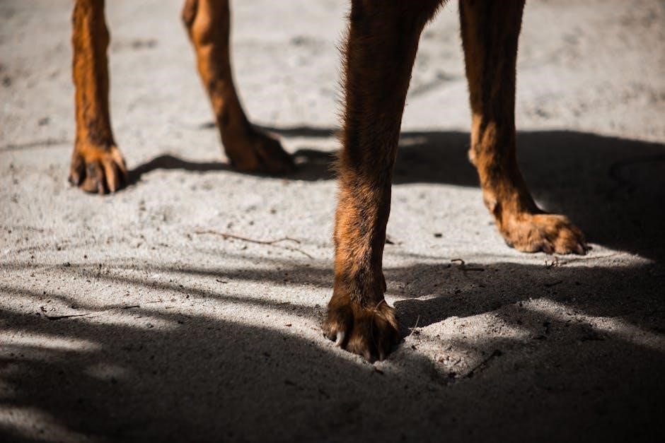
D. Fetlock and Hoof
The fetlock joint, analogous to a human ankle, is a critical articulation connecting the cannon bone to the pastern. Its flexibility absorbs shock during movement and contributes to the cow’s gait. Careful examination of the fetlock reveals any swelling, heat, or abnormal angles, potentially indicating joint issues or injuries.
Distally, the hoof—a keratinous structure—provides protection and traction. Regular hoof trimming is essential to maintain proper balance and prevent lameness. Inspecting the hoof for cracks, bruising, or infections is crucial for overall health. The digital cushion within the hoof contributes to shock absorption. A healthy fetlock and well-maintained hoof are paramount for comfortable and efficient locomotion in cattle.

VI. The Hindquarters: Legs and Feet
The hind legs provide primary propulsion, featuring the thigh, hock, lower leg, and hoof; structural integrity and proper conformation are essential for mobility.
A. Thigh and Upper Hind Leg
The thigh represents the largest muscular mass of the hind leg, providing substantial power for locomotion and supporting the cow’s weight. Its external features include a pronounced musculature, easily visible beneath the skin, and a relatively smooth contour when the animal is in good condition. Palpation reveals the significant gluteal muscles, crucial for propulsion and maintaining posture.
The upper hind leg seamlessly connects to the pelvis, influencing gait and balance. Observing the thigh’s shape and muscle tone can indicate overall health and nutritional status. A well-developed thigh suggests adequate nutrition and exercise, while a noticeably thin or atrophied thigh may signal underlying health issues or insufficient feed intake. Proper assessment of this region is vital for evaluating a cow’s physical condition and identifying potential problems.
B. Hock and Lower Hind Leg
The hock, analogous to the human ankle, is a prominent joint in the lower hind leg, crucial for weight-bearing and flexibility during movement. Externally, it appears as a noticeable prominence, often covered by a short coat of hair. Below the hock lies the cannon bone, a major weight-bearing structure, followed by the metacarpal region and ultimately, the fetlock joint.
Careful examination of the hock reveals its complex structure and potential for injury. Swelling, heat, or lameness in this area often indicates arthritis or sprains. The lower hind leg’s conformation influences the cow’s gait and ability to navigate various terrains. Assessing the alignment and soundness of the hock and lower leg is essential for evaluating overall mobility and identifying potential orthopedic concerns.
C. Fetlock and Hoof
The fetlock joint, situated immediately above the hoof, is a crucial articulation allowing for shock absorption and flexibility during locomotion. Externally, it’s characterized by a puffiness due to the synovial fluid and supporting ligaments. Below the fetlock lies the hoof, a complex structure composed of keratin, providing protection and traction.
Regular hoof care is paramount for bovine health, preventing lameness and maintaining productivity. The hoof’s external features – the sole, wall, and heel – must be routinely inspected for cracks, abrasions, or signs of infection. Proper trimming ensures correct hoof conformation, distributing weight evenly and minimizing stress on the lower limbs. Neglecting hoof health can lead to significant economic losses due to reduced mobility and compromised welfare.

VII. Reproductive Organs (External View)
External reproductive anatomy differs by sex; cows exhibit a vulva, while bulls display a scrotum, both crucial for reproductive functions and health assessments.
A. Vulva (in Cows)
The vulva represents the external opening of the female reproductive tract in cows, serving as a critical point for breeding, calving, and overall reproductive health monitoring. Its structure includes the labia, which are the fleshy folds surrounding the opening, and the clitoris, a sensitive organ analogous to the male penis.
Variations in vulva conformation can influence breeding success, with a properly formed and functional vulva essential for effective semen deposition. Assessing the vulva’s size, symmetry, and tone is a routine part of reproductive examinations performed by veterinarians. Observing any signs of swelling, discharge, or injury is crucial for early detection of potential issues. Proper hygiene around the vulva is also important to prevent infections and maintain reproductive efficiency within the herd.

B. Scrotum (in Bulls)
The scrotum is the external sac housing the testes in bulls, playing a vital role in regulating testicular temperature – crucial for optimal sperm production. This pendulous structure hangs below the body, maintaining a temperature several degrees cooler than the core body temperature.
The scrotal skin is typically thick and wrinkled, providing protection and surface area for heat dissipation. Assessing the scrotum’s size, shape, and texture is a key component of breeding soundness examinations. Palpation of the testes within the scrotum helps determine their size, consistency, and presence of any abnormalities. Any swelling, lesions, or irregularities warrant veterinary attention, as they can impact fertility and reproductive performance.
VIII. The Tail: Structure and Function
The cow’s tail is a significant extension of the vertebral column, serving multiple crucial functions beyond simply swatting away flies. It’s comprised of vertebrae, muscles, nerves, and a switch of hair at the distal end. Primarily, the tail acts as a vital tool for fly control, protecting the animal from irritating insect bites that can transmit disease and cause distress.
Furthermore, the tail plays a role in balance and communication, conveying mood and social signals within the herd. Observing tail position can indicate a cow’s emotional state – tucked tails often suggest fear or submission, while swishing can signal irritation. Proper tail length and health are essential for a cow’s well-being and productivity.